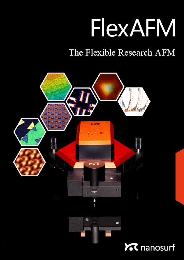
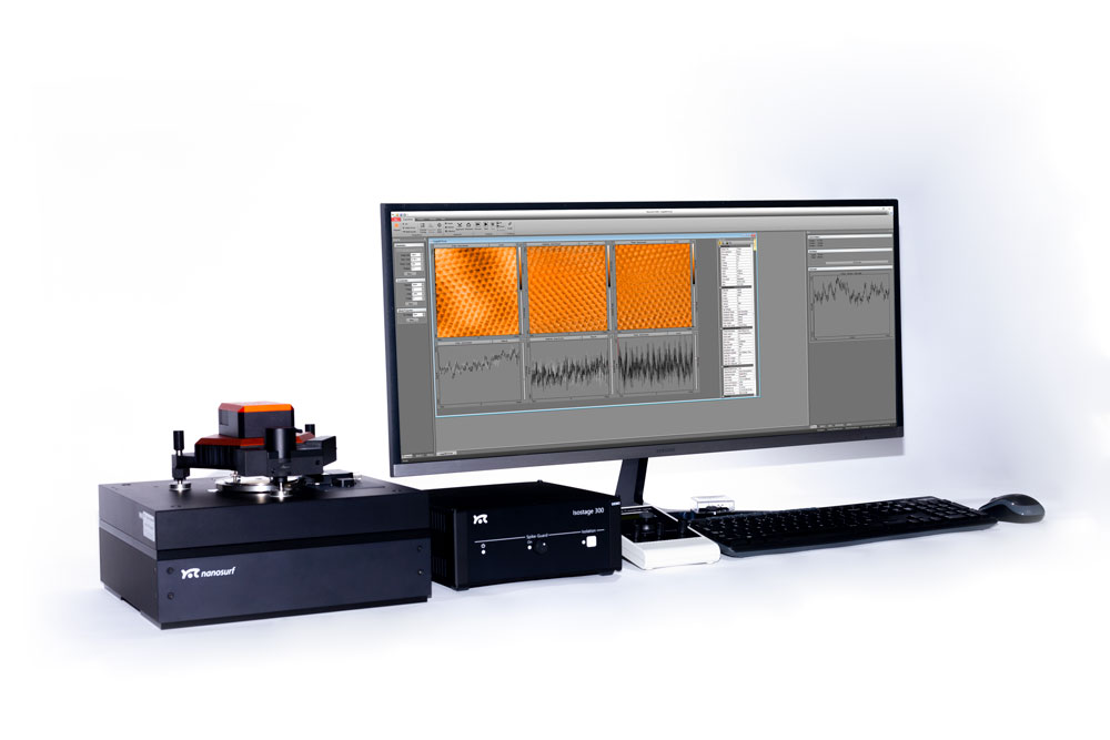
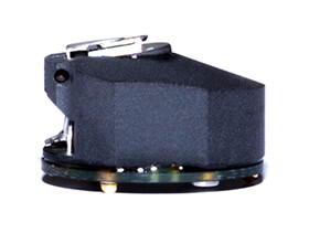
FlexAFM — The flexible research AFM
Overview
FlexAFM — The flexible research AFM
The FlexAFM has been the instrument of choice for hundreds of researchers in need of a reliable and versatile atomic force microscope system. Built with ease of use and configurability in mind, the capabilities of the FlexAFM cover most research needs: Configure your FlexAFM for 2D materials research in a glovebox, or integrate it with your inverted optical microscope for biological applications - no matter what your research goals are, the Nanosurf FlexAFM provides you with an affordable mid-range instrument that you can expand and upgrade as your requirements evolve.
Reliable and robust
Flexure-based scanner provides flat and linear scanning
Versatile
Flexible sample size and weight thanks to tip-scanning technology
Configurable to your needs
From basic research to advanced experiments: Electrical modes; spectroscopy; motorization and automation; inverted optical microscope integration for bio-research and FluidFM; highly suitable for glovebox use
Optional upgrade to CX Controller:
Additional user input/output channels
More flexibility
Performance boost
Future-proof
Compatibility with DriveAFM accessories: Easy upgrade to high-end
User-friendly handling, light and robust design
Download the FlexAFM Brochure
Get a printable PDF of the FlexAFM brochure.

We have been using the FlexAFM system for the last couple of years. The ease of use and its robustness helped us publish a number of articles in high impact journals. I'm very pleased with the performance of your AFM system and would highly recommend your systems for other customers in India and abroad. The technical support from Switzerland as well as local support in India is found to be very satisfactory.
Department of Materials Science & Engineering
INDIAN INSTITUTE OF TECHNOLOGY DELHI
Practical details that really matter in daily use
Cantilever holders with alignment structures are available for use with cantilevers containing alignment grooves. This provides micrometer repositioning accuracy, circumventing laser alignment and allowing you to find the same sample features again and again. Cantilevers enter the image from top to bottom, so sample orientation is always the same, no matter whether you look by eye, CCD camera, or AFM (when scanned at the default scan angle).


FlexAFM imaging modes
This overview shows which modes the instrument is capable of. Some modes may require additional components or software options. For details, please view the brochure or contact us.
Standard imaging modes
Static Force Mode
Lateral Force Mode
Dynamic Force Mode (Tapping Mode)
Phase Imaging Mode
Thermal imaging modes
Scanning Thermal Microscopy (SThM)
Magnetic properties
Magnetic Force Microscopy
Electrical properties
Conductive AFM (C-AFM)
Piezoelectric Force Microscopy (PFM)
Electrostatic Force Microscopy (EFM)
Kelvin Probe Force Microscopy (KPFM)
Scanning Spreading Resistance Microscopy (SSRM)
Scanning Microwave Microscopy (SMM)
Mechanical properties
Force Spectroscopy
Force Modulation
Stiffness and Modulus
Adhesion
Unfolding and Stretching
Force Mapping
Other measurement modes
Lithography and Nanomanipulation
Electrochemical AFM (EC-AFM)
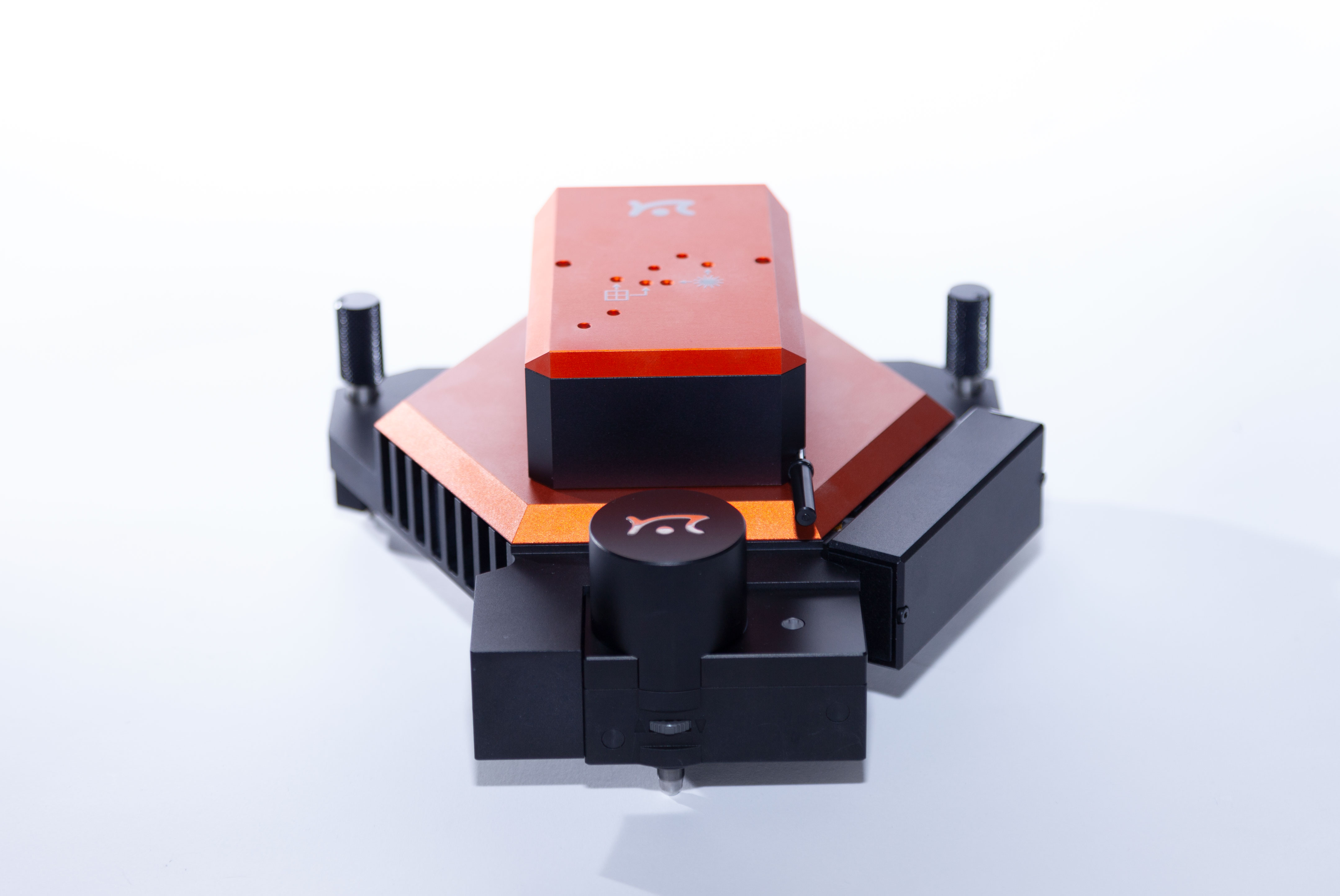
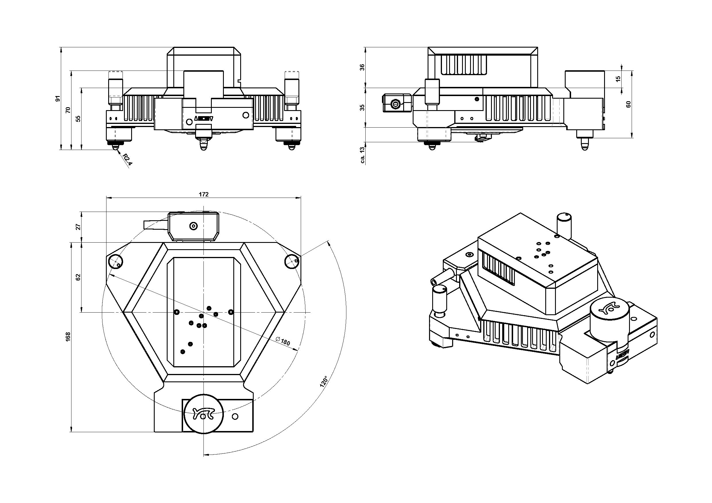
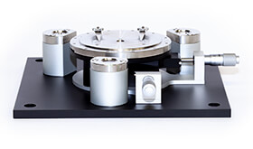
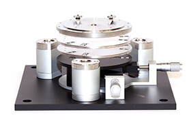
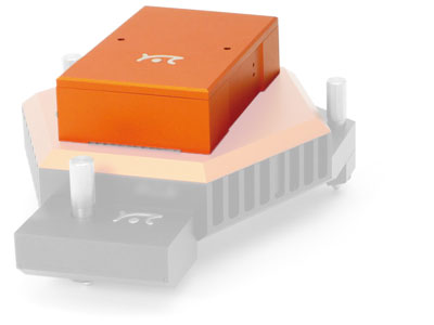
Scan head dimensions*
The FlexAFM scan head has the same footprint as the DriveAFM: it is compatible with all DriveAFM stages.
*since 2023
Life science
Versatility and performance for biology and life science
For success in life science research, scientists depend on professional tools that can readily provide the information needed, regardless of the tasks at hand. By combining key technologies and components, Nanosurf has made the FlexAFM system one of the most versatile and flexible AFM systems ever, allowing a large variety of biological and life science applications to be handled with ease. With the FlexAFM, you can combine the liquid AFM imaging, spectroscopy and nanomanipulation capacity of this system with the high-end optical techniques available for inverted microscopes.
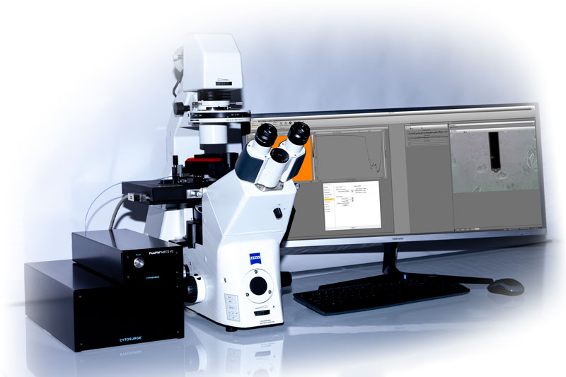
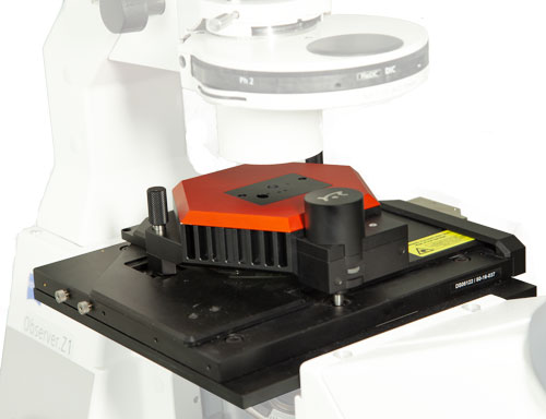
Flexible system design for life science research
FlexAFM comes with manual and motorized stages for seamless integration on Zeiss, Olympus, Nikon and Leica inverted microscopes or with standalone stages. On the inverted microscope, optical and AFM data can be correlated, as shown here for internal limiting membrane (ILM) of the human retina.

The modular stage, cantilever holder, and software concept allows an easy upgrade of the system to access many new possibilities in life science and materials research. Flex-FPM for cell and nano-manipulation, for example, and Flex-ANA for automatic nanomechanical analysis. In addition, advanced modes like MFM and KPFM that were originally developed for the Flex-Axiom system, are also available for FlexAFM. For measurements that don’t need optical access from below, e.g. for the imaging and spectroscopy of samples like bacteriorhodopsin, a standalone stage makes the FlexAFM compatible with the Nanosurf Isostage and Acoustic Enclosure 300, and generally makes the system much more compact.

FlexAFM lifescience application examples
Imaging of type I collagen fibrils
Collagen is the most abundant protein in mammals and contributes to more than 25% of the whole-body protein content. It is the main structural protein of the extracellular matrix of connective tissues and provides e.g. tendons and bone with their tensile strength. Most of the collagen found in mammals is fibrillar type I collagen. Type I collagen fibrils show a typical periodic morphology, the so-called D-banding. D-bands result from staggered self-assembly of individual collagen molecules into larger fibrils with a periodicity of about 67 nm.
Images of collagen fibrils from rat tendon were recorded in Prof. Snedeker's research group at the ETH Zürich. One of the reasearch areas of Prof. Snedeker is tendon mechanics and biology.


The 3D representation of the AFM topography image nicely shows the typical periodic D-banding of type I collagen on all fibrils. The colloagen topography was recorded in static mode using a Nanosensors PPP-XYCONTR cantilever. AFM images were processed using Nanosurf Report Software. Preparation and imaging of collagen fibrils was performed by Massimo Bagnani, Prof. Snedeker research group, Uniklinik Balgrist, Institute for Biomechanics, ETH Zürich, Switzerland.
Measurements on living cultured cells
Mechanobiology is an emerging research area that deals with the effect of changing physical forces or changes in the mechanical properties of cells and tissues. Several diseases, such as fibrosis and atherosclerosis are associated with changes in tissue stiffness. Moreover, in cancer, the metastatic potential of cancer cells depends on their elastic modulus.
Here, the elastic modulus of living cells from a human breast basal epithelial cell line was measured using a Nanosurf FlexAFM system with the Flex-ANA software.


The first image shows the elastic modulus (in kPa) recorded on living breast epithelial cells immersed in cell culture medium. Differences in the elastic modulus within the cell can be clearly observed. The dark area surrounding the cells originate from the much stiffer cell culture dish substrate.
The second image shows the unperturbed cell topography extracted from the force mapping data. The topography is determined from the contact point of each force curve and thus shows the cell topography at zero applied force.


Mapping the elastic modulus data to the 3D topography allows relating the information of both channels. The 3D image was generated using Gwyddion software.
The last image shows the distribution of the elastic moduli extracted from nanomechanical force mapping experiments. The peak at lower moduli corresponds to the stiffness of the cells. The peak at the right originates from the cell culture substrate and shows much higher elastic moduli.
AFM data courtesy of Philipp Oertle, Biozentrum, University Basel.
Single molecule force spectroscopy of bacteriorhodopsin
The force-distance curve below reports the controlled C-terminal unfolding of a single bacteriorhodopsin (BR) membrane protein from its native environment, the purple membrane from Halobacterium salinarium.
Solid and dashed orange lines represent the WLC curves corresponding to the major and minor unfolding peaks observed upon unfolding BR, respectively. The contour length of the stretched polypeptides of the major unfolding peaks is given in amino acids (aa).

This data was recorded using a FlexAFM scan head (10-µm; version 3) in combination with the C3000 controller and a cantilever with a nominal spring constant of 0.1 N/m (Uniqprobe, qp-CONT, Nanosensors).
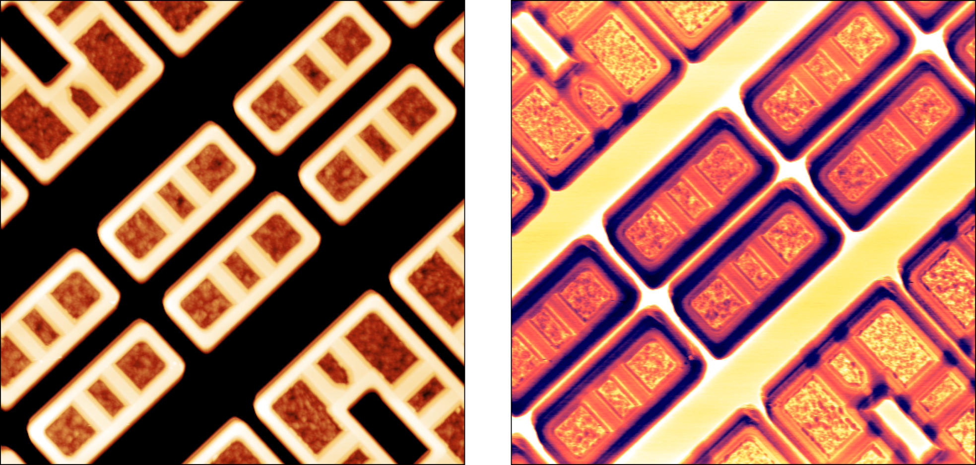
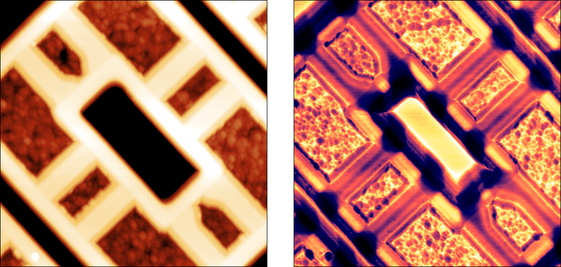
FlexAFM image gallery
")

 for clarity.")
. The deflection image was recorded with a Nanosurf FlexAFM installed on a Zeiss Axiovert inverted microscope. Details of the cell's cytoskeleton are readily discernible through the cell membrane using AFM.")

adsorbed onto muscovite mica (scan size: 2 µm, height range: 1.4 nm).")
")
.")
")
.")
Materials science
The most flexible atomic force microscope for materials research
For success in materials research studies, scientists depend on professional tools that can readily provide the information needed, regardless of the tasks at hand. By advancing key technologies and designs, Nanosurf has made the FlexAFM one of the most versatile and flexible AFMs ever, allowing a large variety of materials research applications to be handled with ease. In combination with the powerful C3000i controller, complex material characterizations are possible.
"The FlexAFM has given us a streamlined and consistent method to cut graphene and graphite gates with ultra-fine precision, allowing for entirely new types of heterostructure devices. What's most remarkable is that this functionality is built in, and is much more simple to setup and use compared to similar techniques in other AFM systems with a much steeper price tag."
J. Ehrets, Kim Lab, Harvard University
The precision and performance you need for your research
The FlexAFM uses an extremely linear electromagnetic scanner for XY movement. This scanner delivers an average linearity deviation of less than 0.1% over the full scan range, top-ranking on the AFM market. The Z-axis is piezo-driven, with a position sensor that enables closed-loop operation. A sensitive cantilever detection system can measure well into the MHz frequency range. The scan head is connected to the full-featured, 24-bit C3000i controller with digital feedback and 2 dual-channel lock-in amplifiers.

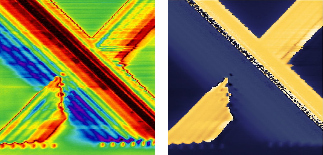
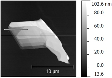
Topography of SrTiO3 in dynamic mode
Strontium titanate (SrTiO3, STO) is an oxide of titanium and strontium exhibiting a perovskite structure. It has interesting and partly unique material properties. It is used as substrate for growth of oxide-based thin films and high-temperature superconductors.
STO forms surfaces that show a layered structure. The thickness of individual layers is in the range of a few Angstrom. Atomic force microscopy is an ideal tool to image and measure these structures.


The sample clearly shows the typical layer structure STO. Here, the layers are not perfectly smooth, but exhibit residual roughness of approx. 125 pm (RMS). This is caused by a non-ideal termination process during the preparation of this STO sample.
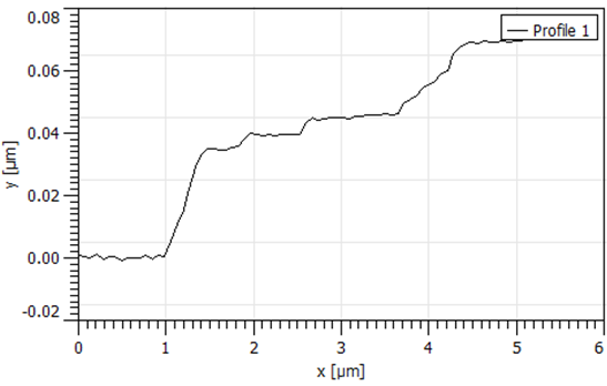
The graph shows the profile of the image shown above along a line extending from the top left to the bottom right corner of the imaged area. The profile also clearly shows the layered structure of the sample and reveals step heights of approx. 4 Å. Similarly in the right panel, the height distribution histogram of the image above clearly shows approx. 4 Å-spaced peaks for the different layers of the sample.
The FlexAFM has given us a streamlined and consistent method to cut graphene and graphite gates with ultra-fine precision, allowing for entirely new types of heterostructure devices. What's most remarkable is that this functionality is built in, and is much more simple to setup and use compared to similar techniques in other AFM systems with a much steeper price tag.
Kim Lab
Harvard University, MA, USA
Topography and KPFM of CVD grown molybdenum disulfide monolayers
In this application note, monolayer MoS2 grown by chemical vapor deposition (CVD) was imaged with Kelvin probe force microscopy (KPFM) using a FlexAFM to study the contact potential difference variation on a single crystal.
Monolayers of MoS2 were grown on a silicon substrate by chemical vapor deposition (Sample courtesy: University of Illinois – Urbana-Champlain).
Non-uniformity of the contact potential signal across the monolayer can inform about doping profiles and other surface defects.


b) Height (top) and KPFM voltage (bottom) profile across monolayer
Measurements using the FlexAFM show a step height of 0.6 nm for the MoS2 monolayer. Concurrent KPFM measurements show a 650 mV contact potential difference between the monolayer and the SiO2 substrate.

All measurements were performed using a FlexAFM system equipped with a ANSCM-PA cantilever from AppNano. Images were processed using MountainsMap SPM.
For more information contact our application scientists
KPFM and MFM on stainless steel
In the experiment shown below, KPFM and topography data were recorded in a single run using a FlexAFM system. Also see the related MFM application.

Scan size: 80 µm x 80 µm
Potential range: 200 mV

Scan size: 80 µm x 80 µm
Height range: 50 nm

Scan size: 80 µm x 80 µm
Phase range: 10°
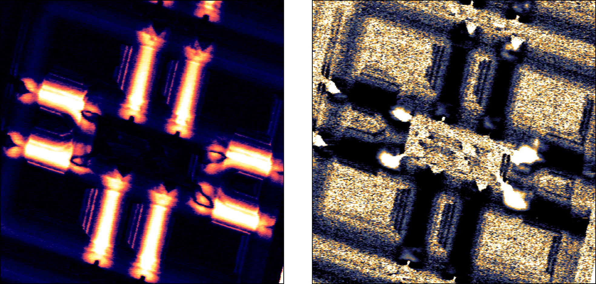
FlexAFM image gallery


")


 on stainless steel (scan size: 80 µm, phase range: 10°)")

")
")
")

")
. The length of the fibers and the size-distribution of the knots can conveniently be analyzed using the measurement tools integrated in the Nanosurf control software.")
")
.")
")
.")
.")
.")
.")
")
Accessories
Modular design: the most flexible system available
With a modular stage concept and various possible cantilever holder types, the FlexAFM is ready for all your current and future needs.

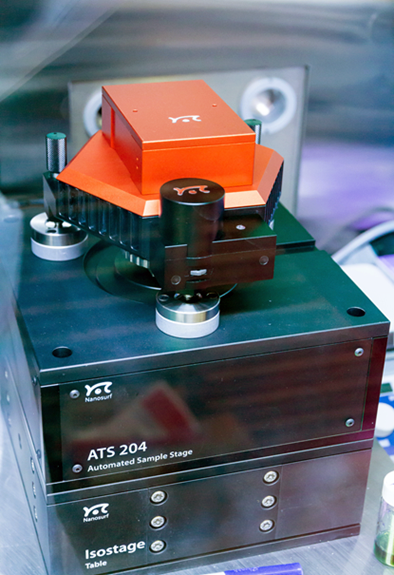
Motorized translation stage 204
- Motorized xyz sample positioning with 32 mm x 32 mm x 5 mm xyz range
- Automated measurements using stage control, scripting and batch manager
Micrometer translation stage (shown on sample stage 204)
Manual sample positioning with 13 mm x 13 mm xy range, 10 μm positioning accuracy
Sample stage for inverted microscopes
- Available for Zeiss, Nikon, Olympus, Leica
- Parallel AFM and optical image axes
Height extension for FlexAFM sample stage
- For measuring higher samples
- Each kit contains spacers to lift the scan head by 8 mm
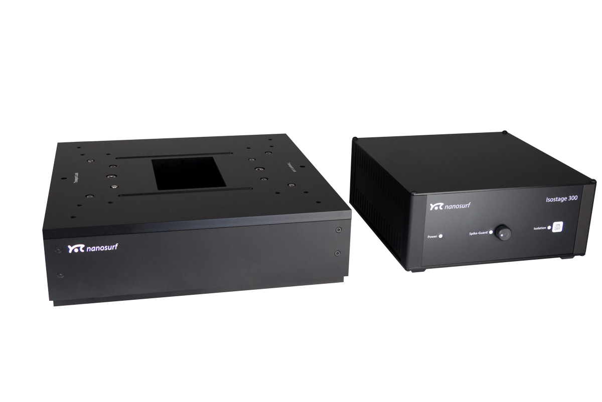
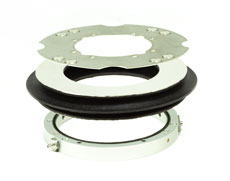
Isostage 300
- Active vibration isolation
- Spike-Guard: detects anomalies and automatically rescans the line
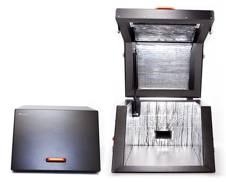
Acoustic Enclosure 350
For use with FlexAFM systems with or without Isostage active vibration isolation table
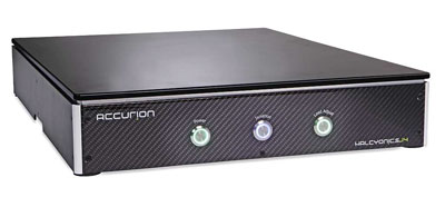
Accurion Halcyonics_i4
Vibration-free measurements on an inverted microscope with the Halcyonics_i4
- State-of-the-art-active vibration isolation system
- Ideal isolation from building vibrations and other disturbances
- Low-profile carbon-design, straightforward handling, easy operation
- Isolation effect starts at 0.6 Hz and achieves max. performance of –40 dB at 10 Hz, where 99.0% of the vibration is isolated
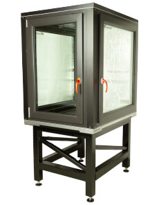
Acoustic enclosure 1100
Protection from airborne noise
- Protect your equipment from airborne noise emitted by air conditioning, venting, door slamming etc.
- Enables you to perform undisturbed experiments with your inverted optical microscope setup.
Cantilever holders
With / without alignment grooves for topography in air and liquid, for electrical measurements, for FluidFM® and for SThM
FlexAFM video camera
- Simultaneous top and side view:
5 MP, 1.5x1.1 mm, color top view and
5 MP, 3.2x3.2 mm, color side view of sample and cantilever - More comfortable sample positioning and approach
Environmental control option
Sample measurements under controlled, dry and/or inert gas atmospheres
FluidFM®
FluidFM®: The established microfluidic tool for single-cell biology and nanomanipulation
FluidFM literally brings microfluidics to the tip of an AFM cantilever. This way, it combines the microfluidics with the force sensitivity and positional accuracy of an AFM allowing a range of exciting applications in single-cell biology and nanoscience.
Nanosurf has a long experience providing AFM systems with FluidFM having launched the FlexAFM with FluidFM in 2011 together with Cytosurge. This integration of FluidFM on Nanosurf platforms has grown over time and now the FluidFM technology is available for Nanosurf's FlexAFM, CoreAFM and DriveAFM platforms.
A tool for research pioneers
- A tool to conduct original research at the frontiers of science from bacterial adhesion to cell-cell interaction and from spotting to injection and extraction.
- A solution with optical, force, and fluidic control.
- FluidFM can also be combined with PicoBalance to measure the mass of cells and microparticles picked up by the FluidFM probe.
FluidFM application examples with FlexAFM
Cell-cell adhesion forces
FluidFM cell adhesion measurements were extended to study cell-cell interaction. This can be the force between a cell (on the cantilever) and a cell below on a substrate (fig. 1 A), but also between a cell and its surrounding cells in a confluent layer (fig. 1 B).
 studied by aspiration of single cells to a hollow FluidFM™ probe (orange). A) Probing the force between a cell immobilized on the cantilever and a cell on the substrate, B) picking a single cell from a confluent layer, probing cell-substrate (purple) and cell-cell (red) interactions.")
Dr. Noa Cohen of Prof. Tanya Konry's group at Northeastern university in Boston studied cell-cell adhesion forces with a Flex-FPM system to gain more insight into tumor progression and metastasis [Cohen et al. (2017)]. Figure 2 shows an optical image of the method depicted in fig. 2 A that was used by Cohen for this study.
 a single cell to be picked up by a FluidFM probe B) the cell aspired to the cantilever and C) the FluidFM probe with aspired cell during a cell-cell adhesion measurement. Data courtesy of Tanya Konry group, Northeastern University, Boston, USA.")
Data courtesy of Tanya Konry group, Northeastern University, Boston, USA.
Single MCF7 breast cancer cell were immobilized on the cantilever. The cell was then pushed on different cell types that were immobilized on a substrate. The measured cell adhesion between MCF7 cancer cells to different cell types were found to develop differently with incubation time. In these experiments, the reversible binding of cells allowed the different cell pairs to be studied with the same probe (fig. 3), allowîng better comparison of measurements.
 Typical force curves between a MCF7 cell aspired to the cantilever and a non-cancerous, fibroblast (HS5) on the substrate at different contact times. B) Development of the force with contact time between the cells. Data courtesy of Tanya Konry group, Northeastern University, Boston, USA.")
Dr. Ana Sancho from the group Prof. Jürgen Groll's group at the University of Würzburg extensively studied the interaction between a cell and its neighbors in a confluent layer of epithelial cells (fig. 2 B) [Sancho et al. (2017)]. Fig. 4 shows the cantilever picking up a cell from a confluent layer (A). After removal the empty space from where the cell was picked up is visible (B). Again, the epthelial cells of interest could be selected based on cell size and cell shape using the inverted microscope.
, Scientific Reports volume 7, 46152.")
Human endothelial cells from the umbilical artery were found to exert strong intercellular forces (figures 6 A & B). The forces could be decreased significantly by overexpression of the so-called Muscle Segment Homeobox 1 (MSX1). The MSX1 induces a endothelial-to-mesenchymal transition. This transition is a process involved in cardiovascular development and disease. Complementary to these adhesion experiments, the Flex-FPM system was also used to perform nano-indentation measurements. To this end colloidal beads aspired to the cantilever and force curves were recorded on the cells.
 Typical single cell force curves of individual cells or cells in a confluent layer, depicting the increase in adhesion force by cell-cell interactions. B) Effect of MSX1 on the observed cell adhesion for individual cells and cells in a monolayer. Grey and black bars: control measurements on individual cells and monolayers, resp., pale and light blue MSX1 treated individual cells and cells in a monolayer, resp. Adapted from: Sancho et al. (2017), Scientific Reports volume 7, 46152.")
Both examples strongly benefitted from FluidFM™ technology provided by the Flex-FPM solution. In case of the confluent layer the large forces of up to over 1.5µN eliminate chemical binding to study cell-cell adhesion forces. In both cases the reversible binding provided the experiments with the necessary speed-up to obtain sufficient statistics.
Spotting and lithography with FluidFM
In cell biology, micropatterning is becoming a widely accepted technique to address cellular behavior at the single-cell level. The ability to guide cell growth by local suppression or stimulation, for example, is beneficial for studying cell growth, for developing cell-based sensors, and for tissue engineering applications. The production of microfabricated biosensors is another application that requires precise placement of biomaterials on a substrate.
FluidFM® technology by Cytosurge enables deposition of (bio)molecules and particles at defined locations with micrometer accuracy and with femtoliter volumes. The closed channel is capable of depositing molecules from a liquid in both air and liquid environment. This enables numerous applications in biomedicine, cell- and microbiology, as well as non-biological nanolithography.


To learn more, also watch the FluidFM webinar presented by Wiley:

Download the FluidFM brochure for more information
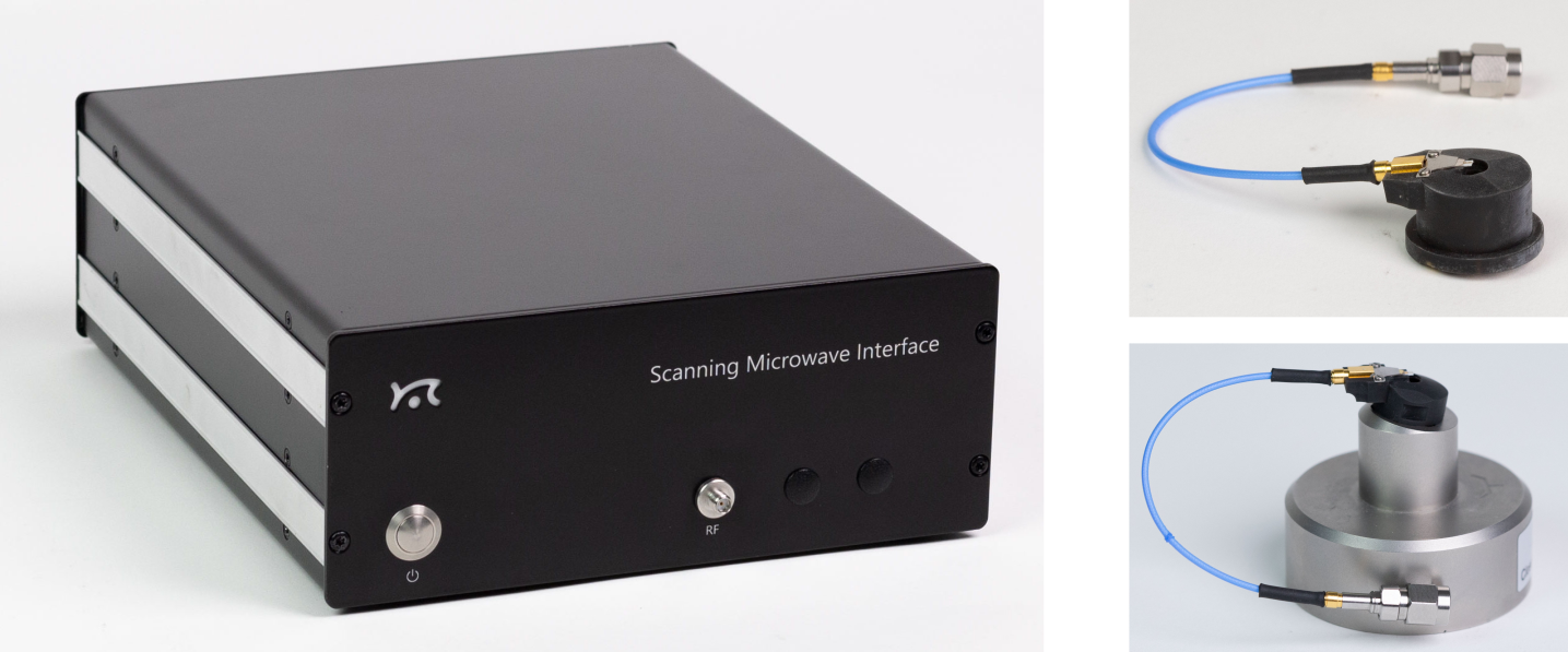
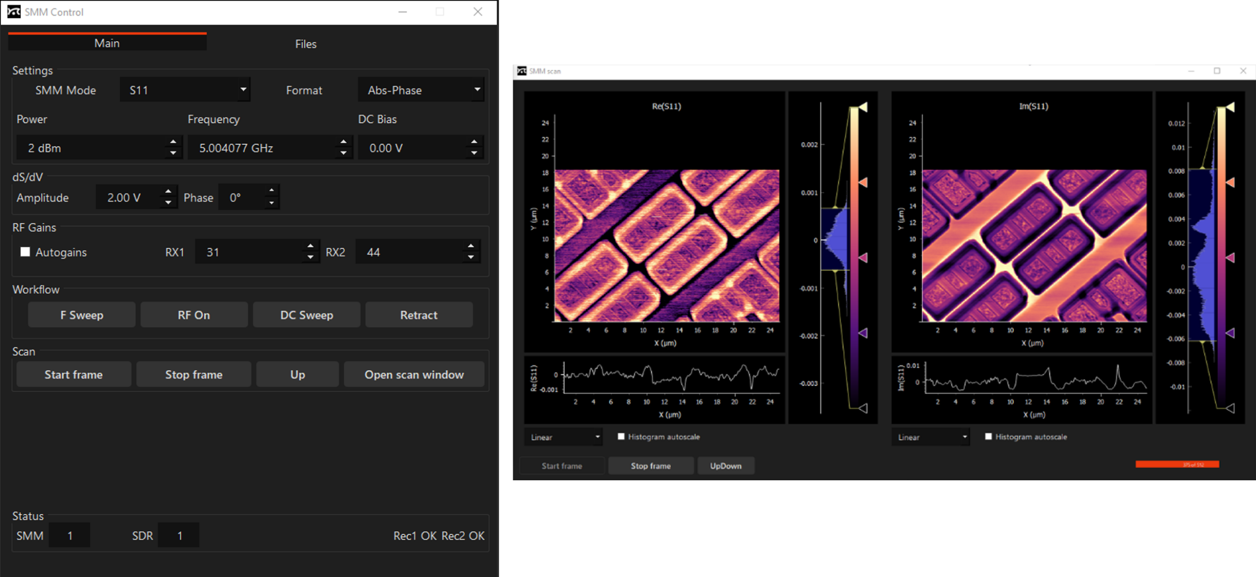
SMM
Scanning Microwave Microscopy (SMM)
Key benefits
- Electrical characterization of materials – dielectrics, semiconductors and metals
- High sensitivity to changes of dopant density in semiconductors
- High sensitivity to DC capacitance
- Minimal sample preparation
- Imaging of buried structures
SMM technique
Scanning microwave microscopy (SMM) is a scanning probe technique, that measures the interactions of a microwave from a sharp tip with the sample. The microwave reflection coefficient (S11 parameter), which is the ratio between the microwave power sent to the tip and the one received back after being reflected at the tip-sample contact, is used to deduce the local tip-sample microwave impedance. The microwave impedance yields information about the local capacitance from which one can deduce the dielectric constant and the dopant density. In terms of the dopant density measurement, the SMM is similar to the scanning capacitance microscopy (SCM), but offers wider range of measurements. SMM can be used for measurements of not only semiconductors, but also dielectric materials and metals, since it is not solely relying of the modulation of the depletion capacitance in the sample.
Understanding the S11 Parameter
The S11 parameter is defined as a function of the complex impedances of reference impedance (Z0) and load (ZL) – Eq.1. Impedance is a complex value and can be represented as a sum of real and imaginary parts – Eq.2. The real part of Z is resistance (R) and the imaginary part can be considered as capacitance (C) -Eq.3. Depending on the sample, resistance and capacitance are functions of conductivity, dielectric constant and carrier density.

Download the SMM brochure
Hardware
Powerful digital signal processing is analysing signal at 1 MHz, far away in frequency from 1/f noise. The signal is digitized early at the analog front-end and such typical issues of analog systems as DC-offset re-adjustment are absent in this configuration. FPGA software has sideband detection capabilities, and dS/dV (dC/dV) measurement does not require any additional hardware, e.g. lock-in amplifiers. Both S11 and dS/dV signals are digital. High working frequency around 5 GHz means better sensitivity to capacitance.

Software
Application examples
SMM provides ultra-sensitive measurements of local capacitances. A capacitance measurement can yield material or device parameters, and it is indispensable for characterization of semiconductor materials, and electronic structures. Read in more detail about application examples in our SMM technical note and application note on Studies of MC2 capacitance standard sample by scanning microwave microscopy (SMM)


Nanosurf SMM Option
The Nanosurf SMM option is currently available for the Nanosurf CoreAFM and FlexAFM and DriveAFM.
| Specifications | |
|---|---|
| Power (dBm) | 4 |
| Frequency (GHz) | around 5.0 |
| DC noise (aF) | 0.5 |
| Doping concentration (1/cm3) | 1015 – 1020 |
| Signal down-conversion | digital |
| Software integration | yes |
| Modes | S11, dS11/dV (dC/dV, dR/dV) without external lock-in |
Glovebox
FlexAFM inside a glovebox
Atomic force microscopy is uniquely suited to measure samples in-situ to monitor real-time, nanoscale surface phenomena. Research in energy storage, catalysis, 2D materials, semiconductors (ALD/MBE), organometallics, photovoltaics, and electrochemistry can often be air-sensitive and require demanding inert gas environments afforded by a glovebox. However, placing an AFM in a glovebox brings challenges such as limited sample access, cumbersome instrument operation, not to mention the additional sources of noise generated by the glovebox that can affect instrument’s performance.
Nanosurf has you covered: our turn-key glovebox AFM solutions offer a low-noise, high-fidelity nanoscale platform coupled with the most stringently controlled environment provided by the glovebox, while also addressing access and ease of use.
Nanosurf has integrated AFMs into a high-performance glovebox by collaborating with MBRAUN, the market leading manufacturer of glovebox workstations. This turn-key glovebox solution means that you don’t have to worry about cutting corners on your AFM measurements to keep your high-purity environment for your experiments.
AFM glovebox workstation package overview
- <0.1ppm O2 and H2O environments for sample preparation and measurements
- Integrated signal feedthrough to enable AFM modes
- Reinforced table & frame to support the AFM and vibration isolation mass
- Vibration dampening to ensure optimal AFM performance
- Large load locks and touch controls to maintain environmental conditions
- Regeneratable oxygen catalyst
- Ease to access, magnetic cantilever holders which are easy to remove and kinematically align upon replacement
- Fully motorized AFM approach and engage
- Motorized sample stages
- Clear, crisp top-down optics for sample alignment (capable of <2µm lateral resolution)


What makes a good AFM for glovebox measurements?
The design and configuration of the AFM is tremendously important to be successful when operating in a glovebox. There is limited space within the glovebox, so a small footprint is desirable. Working with the gloves can be cumbersome, especially manipulating small objects like AFM cantilevers or needing to make multiple, manual adjustments to the AFM to initiate a measurement. Therefore, minimal manual manipulation is ideal to perform AFM for routine measurements. Samples can come in a variety of sizes or substrates, so an AFM capable of accommodating a wide variety of samples without additional preparation simplifies operation of the system.

What makes a good glovebox solution for performing AFM measurements?
An ideal AFM environment is diametrically opposite to that found within a glovebox. Gloveboxes maintain an inert gas environment for work with contamination-sensitive or air-sensitive materials. By design, they precisely control the atmosphere and prioritize design features such as temperature stability, O2 permeability, water content, airlock integrity, and pump performance. The glovebox with all the pumps for atmospheric conditioning introduce vibration, acoustics, and airflow which adversely affect the performance of the AFM. In contrast, an ideal AFM environment is free of excess vibration, has limited airflow, is temperature stable and acoustically quiet over a period of hours. Therefore, careful consideration about instrument compatibility and requirements are needed and not all AFMs are created equal for glovebox work.
Nanosurf's glovebox AFMs...
- ...use a tip-scanner design to ensure the samples are easy to access to load/retrieve regardless of size/shape of substrate (up to 100 mm diameter)
- ...have a compact footprint that saves glovebox real estate.
- ...use a motorized sample engagement and stage translation to allow for the operator to run the AFM without excess ‘hand-in-glove’ time. Once the sample is on the AFM, the entire system is controlled via software from outside the glovebox.
- ...having easy-to-access cantilever holders with alignment grooves facilitates cantilever exchange (FlexAFM) by always positioning the laser in the same place from cantilever to cantilever of the same dimension.
Whether you are looking for a glovebox tool to simply characterize materials in process or your aim is to bring a top-class nanoscale research station into a glovebox environment, Nanosurf’s turn-key glovebox AFM solutions have you covered. The FlexAFM is a capable characterization platform for the budget conscious researcher.
— Dr. Denis Vasyukov, Product Manager Glovebox AFM Solutions, Nanosurf AG
- The inherent compact design saves floorspace without compromising on performance and gives more room in the glovebox for sample preparation and other experiments.
- Samples stages and approach/engagement are motorized, meaning once the sample is loaded on the stage, all control of the AFM happens via software. Nanosurf AFMs are tip scanners which allow for a flexibility for the size/shape of the samples to be measured.
- Alignment grooves in the cantilever holder save you the effort of adjusting the laser between cantilevers.
- There are no small, cumbersome, delicate knobs to turn to load a sample or align the AFM for operation.
What are the applications for a Glovebox AFM?

Any application which requires environmental control, especially of O2 and H2O contamination, will require putting the AFM in a glovebox. Such applications include 2D materials research, semiconductor nanoscale analysis, Lithium (or other Alkali metal) research for energy storage, ALD/MBE materials research, free radical surface work, molecular machines and more.
Nanosurf has a turn-key solution for 2D materials processing in a Glovebox. The FlexAFM-GB is the first AFM specially configured to characterize single-layer 2D material steps within a glovebox. The FlexAFM-GB is guaranteed to be able to resolve step heights ≥200pm within a glovebox at an affordable price. This configuration is ideal for researchers who do not want to expose their materials to the atmosphere for characterization during exfoliation and sample processing, but still need to know dimensions, thickness and basic topography.
Whether you are looking for a glovebox tool to simply to characterize materials in process or your aim is to bring a top-class nanoscale research station into a glovebox environment, Nanosurf can provide you with a suitable turn-key glovebox AFM solution. The FlexAFM-GB is a capable characterization platform for the budget conscious.
What do users of FlexAFM in a glovebox say?
As a long-time Nanosurf customer, I highly value Nanosurf’s exceptionally fast and competent expert-level support. They were of great help in ironing out technical issues with our initial glovebox implementation and responded quickly and positively to all of my requests.

As a long-time Nanosurf customer, I highly value Nanosurf’s exceptionally fast and competent expert-level support. They were of great help in ironing out technical issues with our initial glovebox implementation and responded quickly and positively to all of my requests.
One of the challenges in AFM measurements and especially Kelvin probe force microscopy is the effect of surface contamination. Water films can strongly diminish the feedback behavior of dynamic AFM, inducing tip changes and mode hopping. The surface potential of relatively inert surfaces like graphene is also strongly influenced by the various contaminants present under ambient conditions. The effort to move the experiments to high-resolution AFM systems running in ultra-high vacuum is, however, often too high or even impossible in case of sensitive surfaces.
Therefore, we installed a Nanosurf Flex-Axiom system and an active vibration isolation solution in a N2 glovebox with an O2 and H2O concentration of less than 0.1 ppm. The Flex-Axiom is ideally suited for glovebox operation because of its performance and small footprint, even allowing us to move the AFM in and out of the glovebox through the antechamber without having to disassemble the entire glovebox or disrupt the environmental control of the N2 atmosphere, as would be the case for many other AFM systems.
 and Nanosurf's Marco Portalupi (left) at the Glatzel Lab, discussing the details of the Nanosurf Flex-Axiom AFM inside the glovebox.")


Custom-built, air-tight cable connectors were installed in the side panel of the glovebox, which allows the C3000 controller of the Flex-Axiom system to reside outside of the glovebox, freeing up valuable workspace for experiments and further equipment on the inside. Included in our glovebox setup, for example, is an annealing stage, allowing us to anneal samples inside the controlled environment of the glovebox to get rid of contaminants directly before measurements take place.
After some initial training, tip and sample preparation in the glovebox are easy, and measurement conditions are much improved compared to measurements in ambient. The combination of the Flex-Axiom and a N2 glovebox allows very stable scan conditions of well-defined, clean surfaces. Thus, we have for example been able to perform some intensive research on H- and O-terminated diamond samples, which are strongly affected by H2O adsorption. Details can be found in the following articles:
Silicon-Vacancy Centers in Ultra-Thin Nanocrystalline Diamond Films
S. Stehlik, L. Ondic, M. Varga, J. Fait, A. Artemenko, T. Glatzel, A. Kromka & B. Rezek
Micromachines (2018) 9, 281
Water interaction with hydrogenated and oxidized detonation nanodiamonds — Microscopic and spectroscopic analyses
S. Stehlik, T. Glatzel, V. Pichot, R. Pawlak, E. Meyer, D. Spitzer & B. Rezek
Diamond and Related Materials (2016) 63, 97-102
Read more about glovebox implementations by Nanosurf.
Read more about research at the Nanolino Lab.
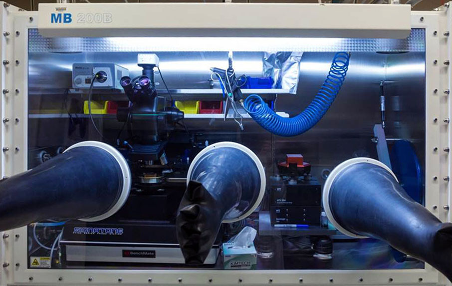
Harvard's Philip Kim investigates mesoscale transport phenomena of low dimensional nanoscale materials with the Nanosurf Flex-Axiom AFM installed in an MBraun Glovebox MB-200B
- The setup allows sample preparation inside the glovebox
- No sample contamination with water or oxygen allows measurement of electrical transport properties
- Cable feedthrough done by MBraun; 2 sets of cables included
- MinusK and IsoStage vibration isolation
Low dimensional materials are of keen interest for many researchers in the materials and physical sciences. These materials have the potential to radically change from the ground up how materials and devices are built and functionalized.
The Kim Laboratory at Harvard University is utilizing the Nanosurf Flex-Axiom atomic force microscope to conduct research on a variety of materials including graphene flakes. The Flex-Axiom AFM offers his group the right combination of performance and ease of use to effectively address and characterize these cutting-edge materials. It also features a footprint small enough to allow passing it through the glovebox airlock, massively saving preparation time when changing the setup in the glovebox.
The Flex-Axiom AFM is installed in an MBraun Glovebox MB-200B, alongside a number of other pieces of equipment including a Signatone Probe Station. With this arrangement, the Nanosurf system is able to effectively operate in an environment maintaining <1ppm O2 and <1ppm H2O.
Software
Control software
The control software for Nanosurf AFMs is an intuitive platform made for performing your AFM measurements efficiently and easily. Our Service team and software engineers are constantly developing and implementing new features and enhancements to further improve the user experience. We regularly publish new and improved versions, which you can download for free. You can install the software on as many computers as you wish to analyze your data.
Free lifetime updates: download all software updates for free
All software updates for Nanosurf control software are free of charge. Our software team is constantly working on new features and improvements to make the user experience better, more intuitive and more efficient.
Software features
Automatic/parameter-free frequency tuning based on cantilever characteristics
Simply choose the cantilever you are using, and the system automatically performs the frequency sweep prior to approaching the sample. No manual setting of parameters is required.
Distance measuring tools: measure the distance between points or lines, the height of features, and more
A selection of different measuring tools allow you to accurately measure angles and distances directly on the acquired measurement image.
Determine the distance between two points or between two parallel lines to make very precise measurement (as shown in the video).

Scripting interface: Python package for COM interface
Python API for data acquisition and control of Nanosurf atomic force microscopes
At Nanosurf we realize that leading researchers are often interested in modifying the standard routines of an instrument, or else everyone would be doing the same thing with the same hardware.
Sometimes there are also application-specific routines, the scope of which we cannot predict and program ourselves. This is why we developed the Nanosurf Scripting Interface.
This interface can be used for automation of routine tasks and for creating new experiments. It gives the users full control over our user interface, and some control over the hardware functionality.
The Nanosurf control software publishes its functionality via the COM interface, by running an instance of COM Automation Server. COM Automation Clients can ask the server during the runtime about its functions and access them. These functions can be accessed through most modern programming languages: Python, C++, C#, VBS, Matlab, JS, LabView, etc.
For ease of use, Nanosurf has created a Python API for its COM interface. We chose Python as our API language, because of its ease of use and learning, popularity and universality, and because of number of data and image processing libraries used in academia and industry.
The Nanosurf Python package can be downloaded and installed from PyPI using pip:
pip install nanosurf
Nanosurf Python Package
See how to take control over the GUI settings in Python. The video shows the most basic actions, like changing the imaging mode, choosing the right cantilever, adjusting image parameters and the PID settings. See how to automatically find the working frequency, start the approach, start scanning, and save the data.

On-demand webinar on Nanosurf's Python API
In this webinar we introduce the Nanosurf Python API and learn how to control Nanosurf instruments with it. We also briefly touch on the processing of the data, stored in NID files, and look at how to create custom user interfaces.
Additional usability features
- Spectroscopy wizard: follow easy steps to set up spectroscopy measurements
- One software UI for all scan heads: no additional learning curve if you use multiple Nanosurf AFM systems
- Automated deflection calibration
- UI layouts for beginners and advanced users
- Highly configurable graph area with mode-dependent auto-layout: the software automatically shows the relevant graphs and information
- Easy file handling with comfort features: auto apply naming conventions, Windows Explorer integration, image gallery, bulk renaming.