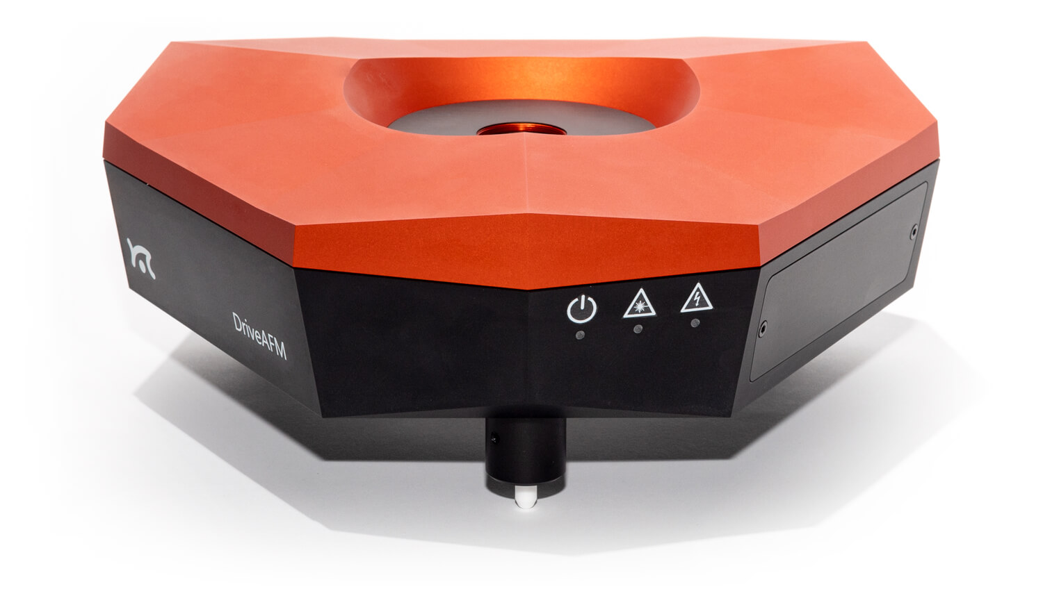
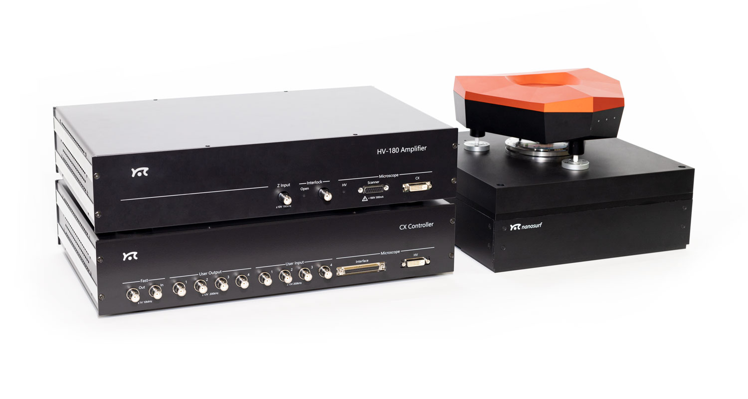
The DriveAFM, Nanosurf‘s new flagship instrument, utilizes the latest technology to deliver stable, high-end performance. It was designed to fulfill the needs of top notch research, today and in the future.
CleanDrive: stable excitation in air and liquid Ultra-low noise Direct drive: high-resolution imaging and large scan area Fully motorized system: full control via software
CleanDrive: stability in air and liquid with photothermal excitation
Photothermal excitation of the cantilever provides unparalleled stability, a linear frequency response, and a high excitation bandwidth in air and liquid environments. These benefits allow measurements at multiple frequencies and highspeed applications and open new horizons for innovative new measurement modes (e.g. Cytomass Monitor).
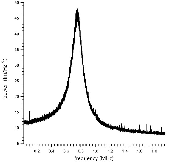
These advantages are amplified in liquids since only the cantilever beam is excited and the liquid environment remains largely unperturbed. This results in clean resonance peaks and not the “forest of peaks” commonly seen with the piezo acoustic excitation of cantilevers. Furthermore, this method of exciting the cantilever is insensitive to changes in the environment and distance to the sample, making the whole measurement system much more stable.
The DriveAFM, with its small light spots, is compatible with the use of small cantilevers, which have several advantages that make them superior in performance. While they have the same spring constant as a conventional cantilever, small cantilevers show a significantly higher resonance frequency and operational bandwidth. Also, the noise performance is better. Due to the small dimensions, the sensitivity is increased, and hydrodynamic drag is decreased. All of this results in better imaging performance.
Photothermal excitation of the cantilever is the new best practice for high-performance AFM systems. The animation shows how the cantilever is actuated by an oscillating laser hitting the base of the cantilever beam.
Ultra-low noise
The DriveAFM has a very low overall noise floor, which is achieved through a combination of a low-noise/low-coherence superluminescent diode and a low-noise/high-bandwidth photodetector used in the beam deflection detection module and the low-noise/high-bandwidth CX Controller. This is the basis for the stable, sensitive, and high-resolution imaging and force spectroscopy capability of the DriveAFM.
Direct drive scanner
The DriveAFM exploits the power of direct drive piezo actuation. The 1:1, non-geared actuation scheme of the
DriveAFM’s flexure scanner provides more force and can drive stiffer scanners. The resulting higher resonance frequency of the scanner components allows for a higher available actuation bandwidth than with geared drives of the same scan size. The direct drive scanner actuation in combination with the low noise 28-bit CX Controller allows for both imaging at large scales and at high resolution. The DriveAFM is the perfect solution for high-resolution imaging of demanding samples such as nanostructures, proteins, or polymeric structures (e.g. DNA), and also for larger, micrometer-sized structures.
Full motorization
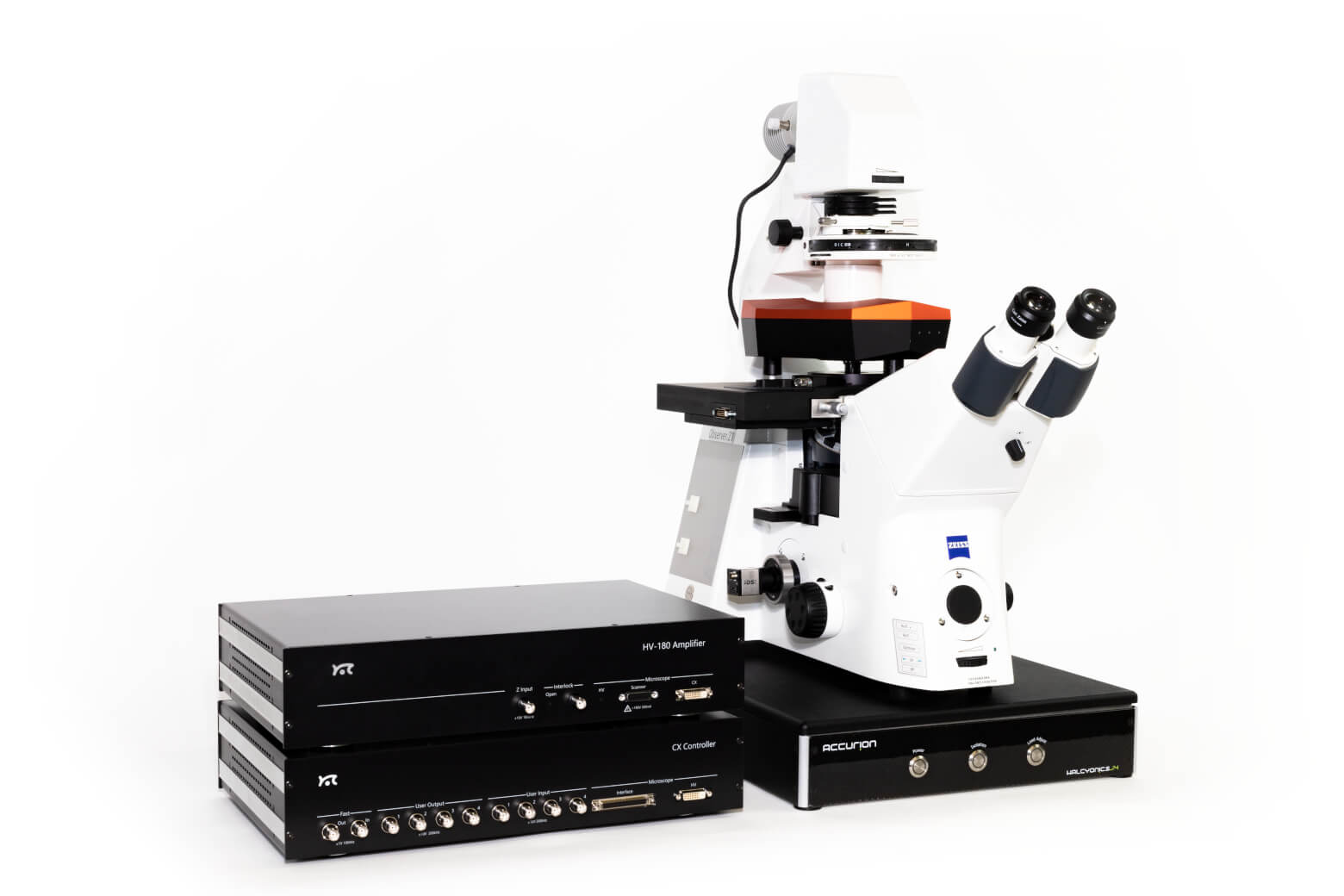
The DriveAFM is the first fully motorized AFM system that can be integrated with an inverted optical microscope. The adjustment of the two light sources for the beam deflection detection system and the CleanDrive photothermal excitation, as well as the photodetector, are fully motorized and can be controlled from the software. The tip approach to the sample is also motorized. The full motorization not only contributes to the ease of use but also allows new possibilities to fully automate the system.
Life science
DriveAFM for biology and life sciences
The DriveAFM plays out all its advantages when it comes to biological applications. The full motorization allows adjusting the lasers and photodetector, and navigating the sample, without interfering with a temperature-controlled environment. CleanDrive excitation provides reliable and clean cantilever tuning in liquid environments. The insensitivity of the CleanDrive towards environmental changes and the sensitivity of small cantilevers facilitate imaging of delicate samples over long periods of time with ease.
Seamless integration of the DriveAFM with an inverted optical microscope allows transmitted light and fluorescence microscopy to be combined with AFM imaging and force spectroscopy. The light sources‘ wavelengths used for CleanDrive (785 nm) and the deflection detection (840 nm) were selected to avoid interference with biological samples and to make fluorescence imaging possible.
The DriveAFM comes with a new line of accessories for biological applications from single-molecule investigations to live cell observations. It includes the new Petri dish holder and the new 150 µm z-actuator, which is essential for cell adhesion experiments. Both sample holders are designed to maintain biological samples at physiological temperature and to be ultimately converted into 2-chamber cell incubators.
The DriveAFM is ready for research that goes beyond conventional imaging. FluidFM®, Cytomass and ANA expand the capabilities of the DriveAFM with multifunctional hollow cantilevers, cell mass measurements and nanomechanical measurement automation.
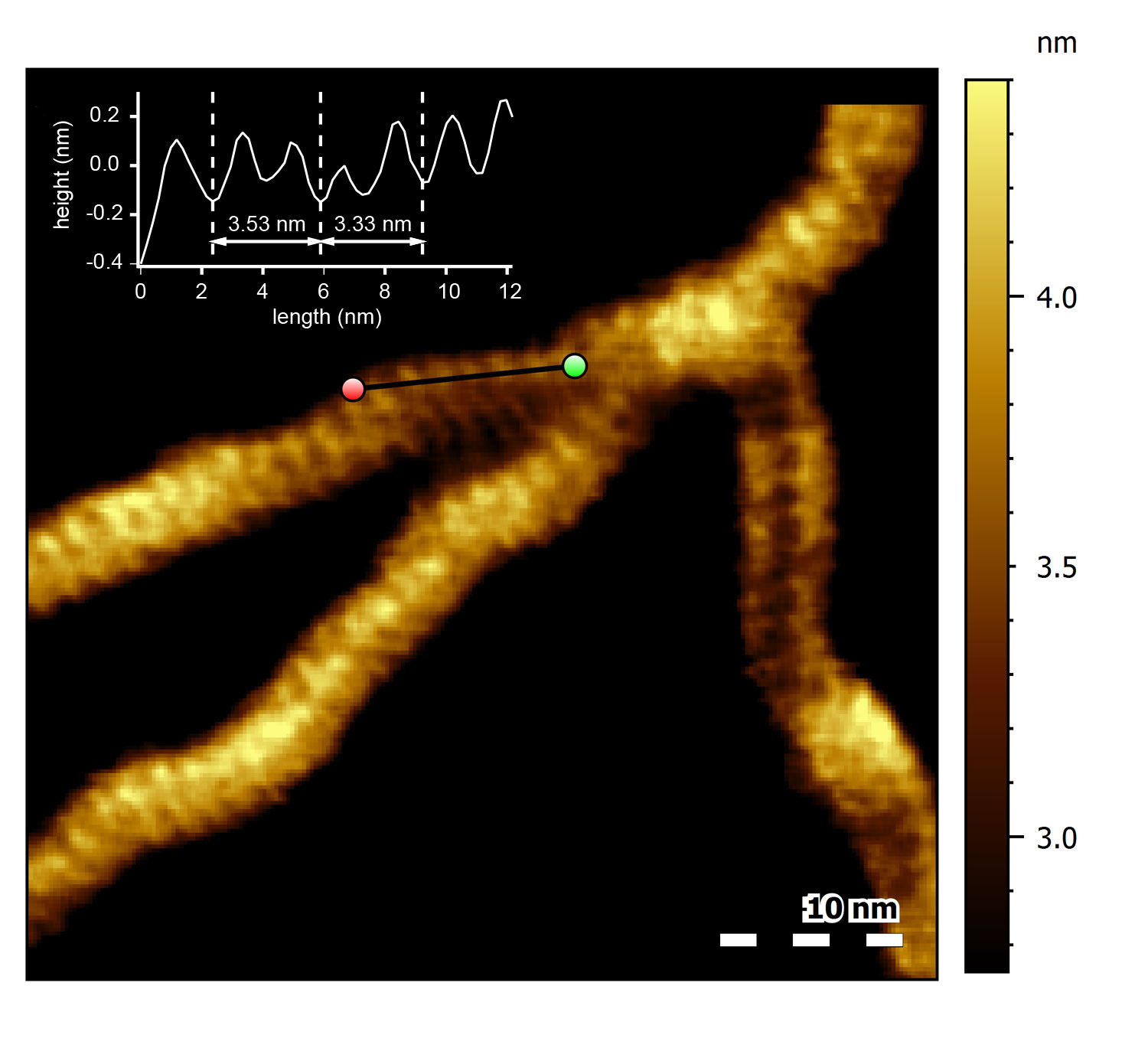
DNA major and minor grooves
High-resolution topograph of double-stranded DNA (dsDNA) adsorbed to mica in buffer solution. Several dsDNA strands can be observed. All of them show a characteristic periodic pattern. Click on the image for a close-up: the black line indicates the location of the cross section shown in the inset. The average spacing between every second-next groove in the section corresponds to 3.4 nm, the characteristic pitch distance of a helical turn of B-DNA. The valleys in the section correspond to the major and minor grooves found in dsDNA.
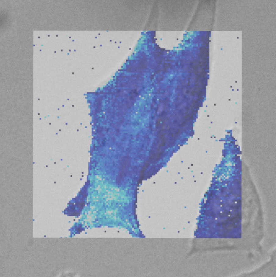
Force map of live fibroblast cell
Force map of live fibroblast cell in cell culture medium at 37°C. The force map is superimposed on the DIC image of the cell.
Seamless integration of the DriveAFM with inverted optical microscopes allows transmitted light and fluorescence microscopy to be combined with AFM imaging and force spectroscopy. The wavelengths of the light sources used for CleanDrive (785 nm) and deflection detection (840 nm) were selected not to interfere with biological samples and fluorescence imaging.
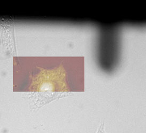
Colocalization phase image
AFM image superimposed on the optical DIC image of the cell.
FluidFM® ready, Cytomass ready, ANA ready
The DriveAFM is ready for research that goes beyond conventional imaging. FluidFM, Cytomass and ANA expand the functionality of the DriveAFM with multifunctional hollow cantilever, cell mass measurements and measurement automation.
Materials science
DriveAFM for materials science
The DriveAFM combines performance and a wide range of applications important for material science research. Its unique direct drive tip scanner technology paired with CleanDrive are key for fast and stable operation in air and liquid. The tip scanner design makes the performance of the DriveAFM independent of the mass of the sample under investigation also allowing measurement on heavy samples. The full motorization not only simplifies working with the system but also facilitates automated measurements addressing different areas of a sample.
Besides reliable topographic imaging, the DriveAFM also features a complete set of different modes to investigate the nanoelectrical (e.g. C-AFM, KPFM, or PFM) or nanomechanical properties of your sample. The universe of accessories available for the DriveAFM offers extended functionality such as heating or cooling the sample, applying a variable magnetic field, detecting low electrical currents or investigating with in situ AFM imaging the changes taking place on electrodes during electrochemical processes.
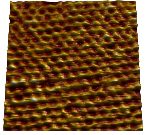
Mica
Atomic lattice of mica imaged in air.
Image size: 7 nm.
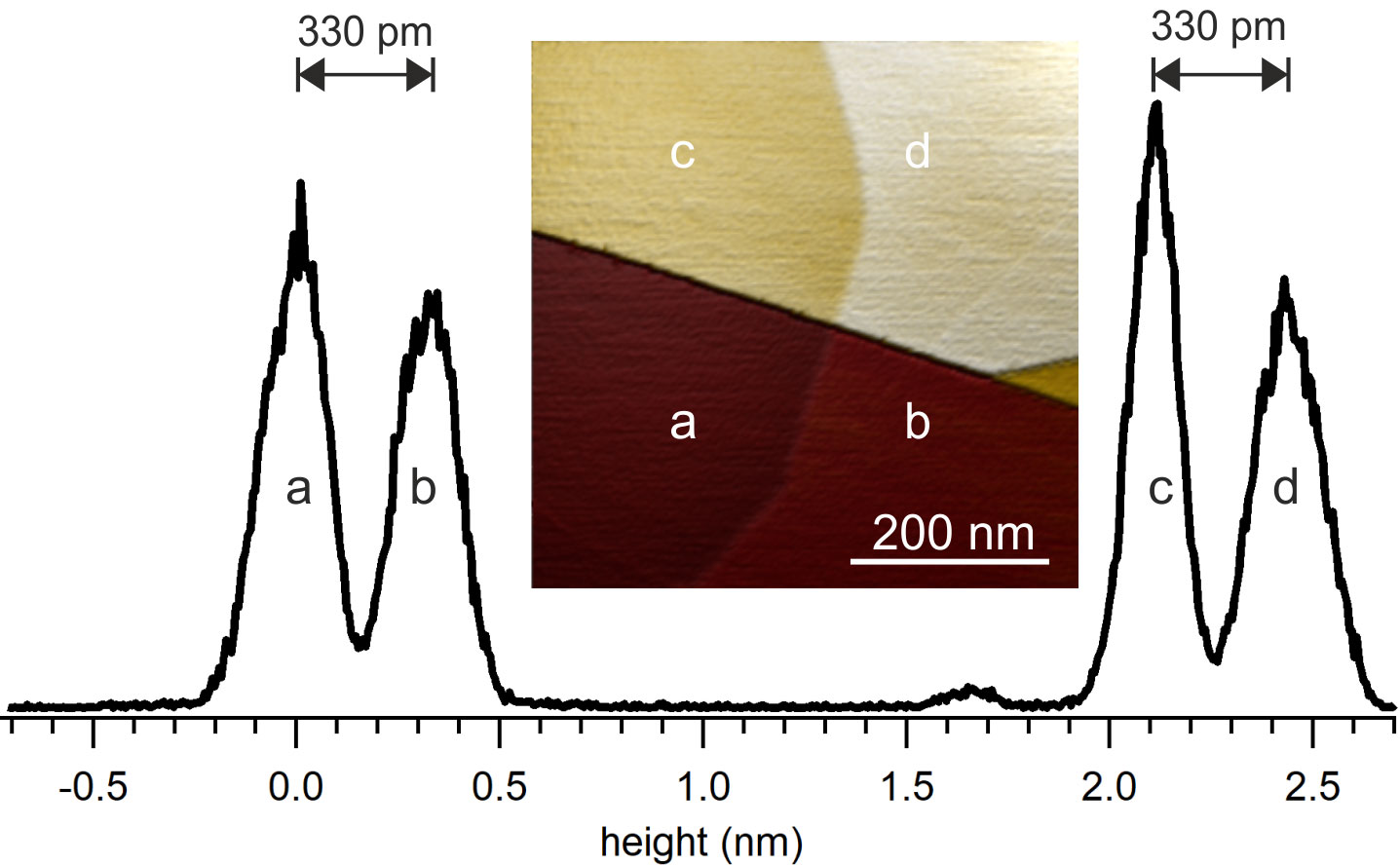
Topography of an HOPG surface imaged in air
Topography of an HOPG surface imaged in air: the surface shows different steps between different graphite layers. Height histogram of the HOPG surface shown on the right. Image size: 500 nm. The spacing of two neighboring peaks in the histogram corresponds to 330 pm, the expected height of a graphite layer.
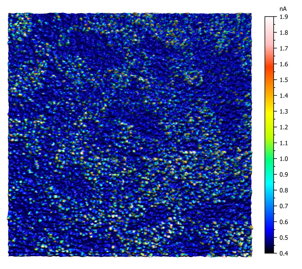
Conductive AFM on ITO
This conductive AFM measurement on ITO reveals the heterogeneous conductivity of an untreated ITO surface. The image was recorded using an EFM cantilever with a tip bias of 5mV. The current was measured using the C-AFM sample holder. Color scale 1.5 nA.
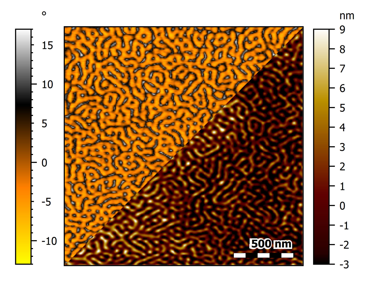
PS-Pb-PS triblock copolymer thin film on mica imaged at 20 Hz line rate
This image shows the phase (top) and topography (bottom) of an unannealed PS-PB-PS triblock copolymer thin film on mica imaged at 20 Hz line rate using an USC-F1.2-k7.3 cantilever.
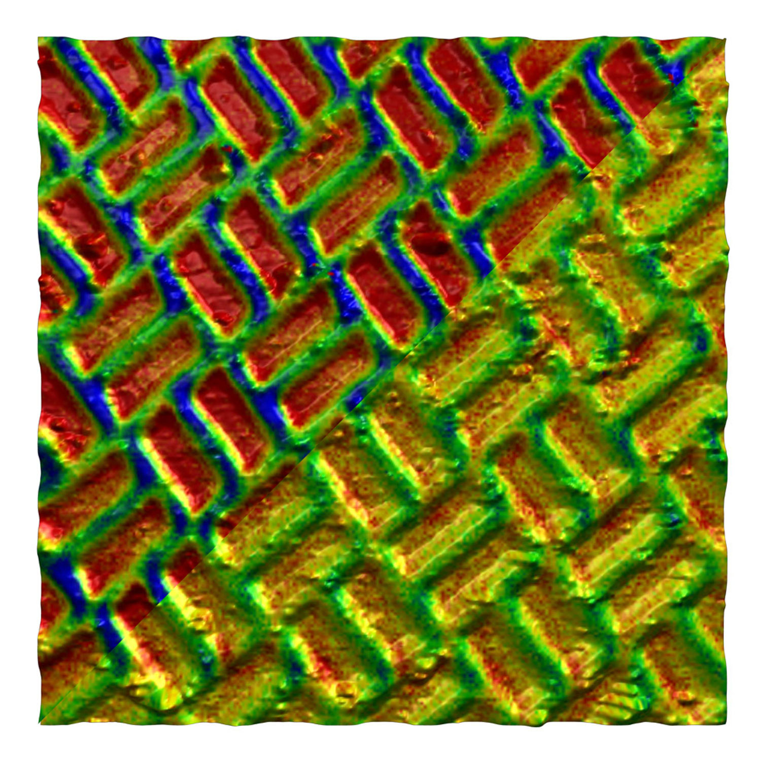
MFM images of a Shakti lattice
MFM images of a Shakti lattice while applying different in-plane magnetic fields (top: 200 mT, bottom: 50 mT). The images represent the 3D topography of the lattice, and the color scale overlaid corresponds to phase shift induced by magnetic interactions. Image size: 8 µm, phase signal range: 1°.
Versatility without compromise
The DriveAFM offers a wide range of applications to investigate material properties. Besides pure topographic imaging, the DriveAFM also features a complete set of different modes to investigate the nanoelectrical (e.g. C-AFM, KPFM, or PFM) or nanomechanical properties of your sample. The universe of accessories available for the DriveAFM offers extended functionality such as heating or cooling the sample, applying a variable magnetic field, detecting low electrical currents or investigating with in situ AFM imaging the changes taking place on electrodes during electrochemical processes.
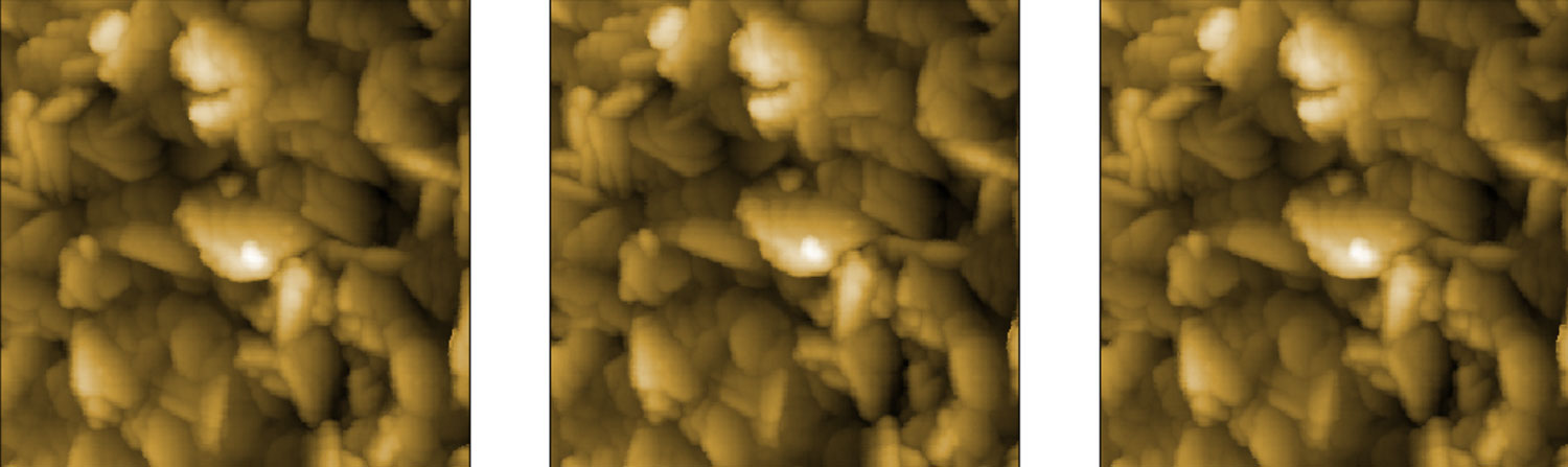
Consecutive unattended imaging of a TipCheck sample with CleanDrive excitation. After 100 images no noticeable change in the tip curvature could be observed. Measurement of tip-check sample over 10 hours. 1st, 50th and 100th image.
Specifications
DriveAFM imaging modes
This overview shows which modes the instrument is capable of. Some modes may require additional components or software options. For details, please view the brochure or contact us directly.
Standard imaging modes
Static Force Mode
Lateral Force Mode
Dynamic Force Mode (Tapping Mode)
Phase Imaging Mode
Magnetic properties
Magnetic Force Microscopy
Electrical properties
Conductive AFM (C-AFM)
Piezoelectric Force Microscopy (PFM)
Electrostatic Force Microscopy (EFM)
Kelvin Probe Force Microscopy (KPFM)
Mechanical properties
Force Spectroscopy
Force Modulation
Stiffness and Modulus
Adhesion
Unfolding and Stretching
Force Mapping
Lithography and Nanomanipulation
Electrochemical AFM (EC-AFM)
System specifications
Scan head
Scan size
typ. typ. 100 µm x 100 µm x 20 µm
min. 95 µm x 95 µm x 18 µm
Read-out light source
850 nm low-coherence SLD
CleanDrive light source
785 nm laser
Photodetector bandwidth
≥8 MHz
Standard / maximum sample size
100 mm / 150 mm
Z-height noise dynamic
<30 pm (RMS)
Z-height noise static
<30 pm (RMS)
DC detector noise*
<5 pm (RMS, 0.1 Hz – 10 kHz)
AC detector noise*
<25 fm/√(Hz) above 100 kHz
Approach
10 mm motorized, parallel
(*) measured with a USC-F1.2-k7.3 cantilever
CX Controller specifications
High resolution outputs (DAC)
12x 28 bit, 1 MHz/sampling; thereof 4x user DAC (optional)
Fast outputs (DAC)
4x 16 bit, 100 MHz/sampling; thereof 1x user DAC (optional)
High resolution inputs (ADC)
12x 20 bit, 1 MHz/sampling; thereof 4x user ADC (optional)
Fast inputs (ADC)
3x 16 bit, 100 MHz/sampling; thereof 1x user ADC (optional)
Signal analyzers
2 signal analyzer function blocks that can be configured as dual channel lock-in
FPGA module and embedded processor
System-on-chip module with low-latency FPGA signal processing at 100MHz and dual-core ARM processor, 2GB RAM, 1.5GHz clock
Scan control
28-bit X/Y/Z-DAC
Detector inputs
Deflection/lateral signals each 20 bit
Digital sync, Spike-Guard
2-bit line/frame sync out 5 V/TTL galvanically isolated, Spike-Guard input
Clock sync
10MHz/3V clock input to synchronize data acquisition and processing