Performance without compromise
- Home

- FluidFM Tab
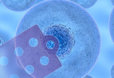
FluidFM
The established microfluidic tool for nanomanipulation and single-cell biology
FluidFM probe microscopy (FPM) combines the force sensitivity and positional accuracy of an AFM with FluidFM technology by Cytosurge to allow a whole range of exciting applications in single-cell biology and nanoscience.
As Cytosurge's initial cooperation partner for this innovative technology, Nanosurf has the longest experience providing AFM systems with the FluidFM® add-on, — launching FluidFM® technology for its AFMs as early as 2013.
FluidFM add-ons are currently available for the DriveAFM, FlexAFM and CoreAFM platforms, and a unique integrated FPM solution exists for the FlexAFM.
Highly accurate pressure, force, and position control with optical sample access
- Fully integrated system with user-friendly FluidFM® ARYA operator software
- FluidFM® microfluidics control system
- Compatible with major inverted microscope brands
Different FluidFM® probes: hollow cantilevers designed for specific applications
- FluidFM® micropipettes: tipless cantilevers with opening at the cantilever end
- FluidFM® nanopipettes: cantilevers with opening at the tip apex
- FluidFM® rapid prototyping probes: cantilevers with closed pyramidal tips, ready for FIB milling
Pioneering research within reach
- A tool to conduct original research at the frontiers of science
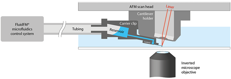
An integrated solution with optical, force, and fluidic control


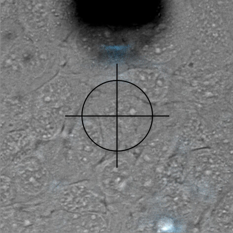
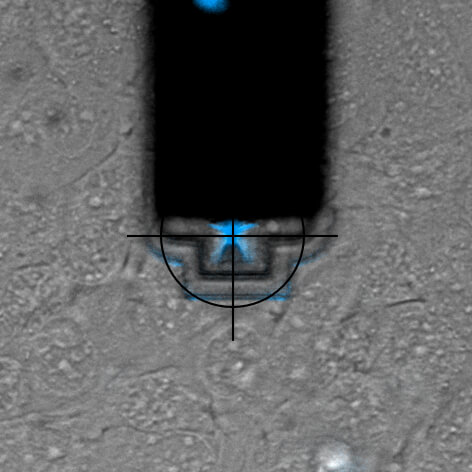
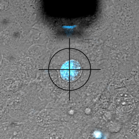
Nuclear injection
DAPI is a fluorescent marker that binds into the minor groove of DNA. Since a cell's DNA is present in the nucleus, it is a commonly used nuclear stain agent. Before conventional labelling, cells are fixed and the membrane permeabilized to allow the dye to enter the cells. Due to the high required concentration, it is conventionally not used for living cells. In the experiment below, FluidFM is used to directly inject the nucleus of living cells with DAPI. DAPI's excitation maximum is at 358 nm and its emission is in the blue part of the light spectrum (461 nm). Sample courtesy: Dr. Nico Strohmeyer, Biophysics group, Prof. D.J. Müller, ETH Zurich
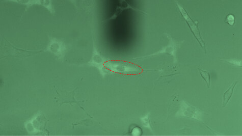
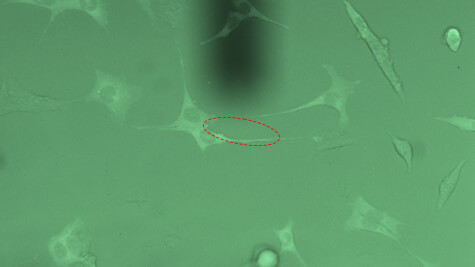
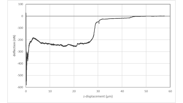
Single-cell force-spectroscopy
Single-cell force-spectroscopy studies are genarally hampered by the relatively slow pace at which a statistically relevant amount of data can be obtained. FluidFM® can dramatically increase the amount of data that can be recorded in a single day: By applying underpressure to the system, FluidFM® cantilevers can attach to a selected cell within seconds. Likewise, the probe can be immediately re-used after each adhesion experiment by releasing the detached cell from the probe through the application of overpressure. The maximum cell adhesion forces that can be measured using FluidFM are in the range of 500 pN to 1600 nN. The latter value represents a one-order of magnitude increase compared to conventional AFM approaches.
Innovative and intuitive handling
With its touchscreen interface and predefined experimental workflows, e.g for spotting and single-cell injection and extraction, or for bacterial and cell adhesion force measurements, the intuitive FluidFM operator software will guide you through each of your experiments step-by-step.
FluidFM opens the door to many fascinating new experiments


To learn more, also watch the following webinar: