Anal. Chem. 90, 11453–11460 (2018)
Livie Dorwling-Carter, Morteza Aramesh, Hana Han, Tomaso Zambelli, and Dmitry Momotenko*
Summary
Surface charge and surface potential are material characteristics that influence functional properties of surfaces. Kelvin Probe Force Microscopy (KPFM) is conventionally used to study surface charge and surface potential in vacuum and air. However, in liquids with high chemical polarity the surface charge is efficiently shielded by an electrical double layer (EDL), a structure that appears at the interface of a surface exposed to fluid. This EDL hampers KPFM measurements in liquid.


Scanning ion conductive microscopy (SICM) can measure the surface charge of surfaces in aqueous solutions using glass micropipettes. The current through a glass micropipette is not only influenced by the gap size between pipette and surface but also by the charge of the surface. This is due to rectifying properties of the current through small openings caused by the surface charge around these openings. When using the glass micropipette, it is challenging to separate topography from surface charge. At the ETH Zurich, a workaround was found using special AFM probes. Probes used for scanning probe microscopy exists in many different configurations and geometries. The classical ones are either the diving-board or triangular shaped silicon or silicon-nitride cantilevers. In more sophisticated applications such classical probes are replaced with optical fibers or even microfluidic probes! It is the latter – microfluidics-based AFM probes, also referred to as FluidFM® probes – that captured the attention of a team of scientists led by Tomaso Zambelli for many different applications, e.g. injection/extraction, patch clamp or cell adhesion experiments. With Dmitry Momotenko joining the team they pursued studies of surface charge and topography at a sample-electrolyte interface with FluidFM probes (Dorwling-Carter et al., Analytical Chemistry, 2018 DOI: 10.1021/acs.analchem.8b02569).
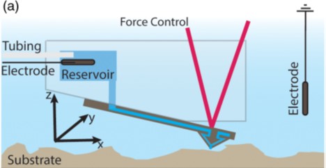
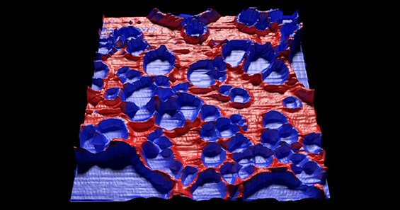
They used a FluidFM® add on from Cytosurge that couples the Nanosurf FlexAFM with SICM using microfluidic cantilevers. A schematic of the setup is shown in fig (a). The FluidFM probes have nanofluidic channels along the length of the cantilever with a 300 nm opening at the tip apex. This method allows simultaneous topographical and current imaging. The topography imaging is done as conventional atomic force microscopy through the deflection of these microfluidic probes. The surface charge information is derived from ion current sensing through the microfluidic cantilever. In this paper, the group reports the results of their investigation of the surface charge density on a partial polystyrene film over a glass substrate as well as a microcontact printed poly-D-lysine film. Figure b shows the overlay of the ion current on surface topography of the polystyrene film. This FluidFM setup provides several significant advantages including increased sensitivity towards lower salt concentrations and higher acquisition rates. The method described in this paper relies on contact mode operation. Expanding to a non-contact or intermittent contact mode will expand such measurements to other materials. In the meantime, this paper demonstrates fast, positionally accurate, simultaneous topographical and charge imaging, providing the KPFM analog for samples immersed in liquid.

