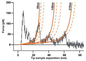
The force-distance curve below reports the controlled C-terminal unfolding of a single bacteriorhodopsin (BR) membrane protein from its native environment, the purple membrane from Halobacterium salinarium.
Solid and dashed orange lines represent the WLC curves corresponding to the major and minor unfolding peaks observed upon unfolding BR, respectively. The contour length of the stretched polypeptides of the major unfolding peaks is given in amino acids (aa).

This data was recorded using a FlexAFM scan head (10-µm; version 3) in combination with the C3000 controller and a cantilever with a nominal spring constant of 0.1 N/m (Uniqprobe, qp-CONT, Nanosensors).
Related content:
Flex-ANA
- Flex-ANA measurement on gelatin hydrogels
- Flex-ANA measurement on medical tubings
- Flex-ANA measurements on living cultured cells
Flex-FPM
View application note (PDF)
