Mechanobiology is an emerging research area that deals with the effect of changing physical forces or changes in the mechanical properties of cells and tissues. Several diseases, such as fibrosis and atherosclerosis are associated with changes in tissue stiffness. Moreover, in cancer, the metastatic potential of cancer cells depends on their elastic modulus. Here, the elastic modulus of living cells from a human breast basal epithelial cell line was mapped.

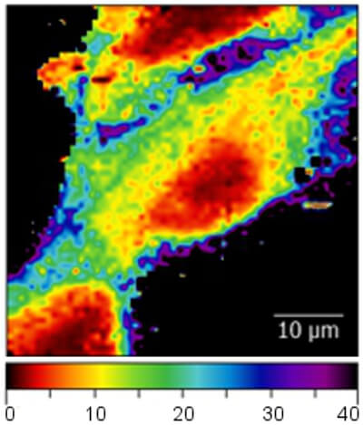
Elastic modulus (in kPa) recorded on living breast epithelial cells immersed in cell culture medium. Differences in the elastic modulus within the cell can be clearly observed
The dark area surrounding the cells originate from the much stiffer cell culture dish substrate.

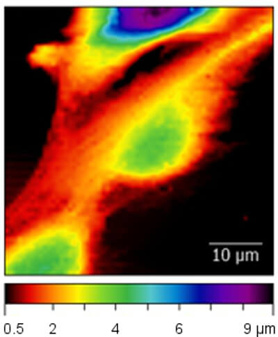
The image shows the unperturbed cell topography extracted from the force mapping data. The topography is determined from the contact point of each force curve and thus shows the cell topography at zero applied force.

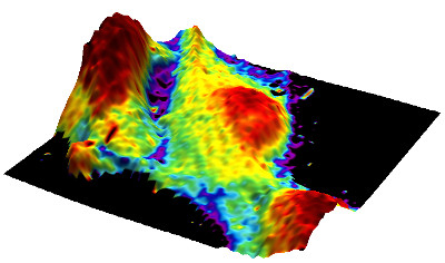
Mapping the elastic modulus data to the 3D topography allows relating the information of both channels.
The 3D image was generated using Gwyddion software.

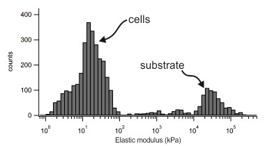
Distribution of the elastic moduli extracted from nanomechanical force mapping experiments. The peak at lower moduli corresponds to the stiffness of the cells. The peak at the right originates from the cell culture substrate and shows much higher elastic moduli.
AFM data courtesy of Philipp Oertle, Biozentrum, University Basel
Nanosurf application note AN00823

