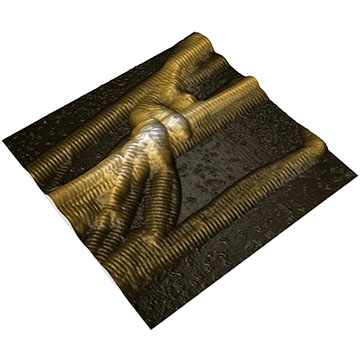
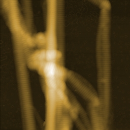
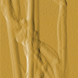
Collagen is the most abundant protein in mammals and contributes to more than 25% of the whole-body protein content. It is the main structural protein of the extracellular matrix of connective tissues and provides e.g. tendons and bone with their tensile strength. Most of the collagen found in mammals is fibrillar type I collagen. Type I collagen fibrils show a typical periodic morphology, the so-called D-banding. D-bands result from staggered self-assembly of individual collagen molecules into larger fibrils with a periodicity of about 67 nm.
Images of collagen fibrils from rat tendon were recorded in Prof. Snedeker's research group at the ETH Zürich. One of the reasearch areas of Prof. Snedeker is tendon mechanics and biology.
View or download application note as PDF

The 3D representation of the AFM topography image nicely shows the typical periodic D-banding of type I collagen on all fibrils.

The colloagen topography was recorded in static mode using a Nanosensors PPP-XYCONTR cantilever.
AFM images were processed using Nanosurf Report Software.

Preparation and imaging of collagen fibrils was performed by Massimo Bagnani, Prof. Snedeker research group, Uniklinik Balgrist, Institute for Biomechanics, ETH Zürich, Switzerland.
Nanosurf application note AN00955

